fNIRS: Reading the Brain with Light
fNIRS shines near-infrared light through the scalp and reads the hemoglobin response beneath — giving researchers a portable, wearable window into cortical activity that works on infants, moving participants, and two people talking to each other. This article explains the physics, the hemodynamic signal, how fNIRS compares to fMRI and EEG, the landmark 1993 motor study by Villringer et al., and what the technique opened up in developmental and social cognitive neuroscience.

In 1977, Frans Jöbsis held a slice of beefsteak up to a bright lamp and noticed something odd: red light passed through it. That observation — published in Science that same year — launched a research program that would eventually put portable brain scanners on the heads of infants, rock climbers, and surgeons. The technology it seeded is functional near-infrared spectroscopy, or fNIRS.
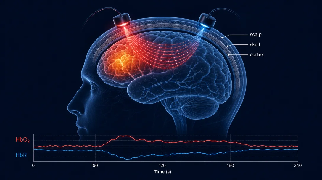
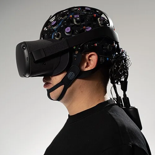
fNIRS does something that sounds almost too simple to work: it shines low-intensity near-infrared light into the scalp, and from the light that scatters back out, it infers where oxygenated blood is flowing in the brain beneath. Compared to fMRI — which requires lying motionless inside a 1.5-tonne magnet — the trade-offs are striking. You lose depth and spatial resolution. You gain a device the size of a bicycle helmet that participants can wear while walking down a corridor, chatting with another person, or sitting in an MRI scanner simultaneously.
That trade-off profile has made fNIRS one of the fastest-growing methods in cognitive neuroscience. The number of peer-reviewed fNIRS papers doubled every 3.5 years through the 2010s, 1 and the Society for fNIRS — founded in 2014 — now holds conferences drawing hundreds of submissions per cycle.
How the physics works
The key is a narrow spectral window between roughly 650 and 950 nm. In this "optical window," skin, fat, and bone absorb relatively little light. Hemoglobin, by contrast, absorbs a lot — and it absorbs differently depending on whether it is carrying oxygen.
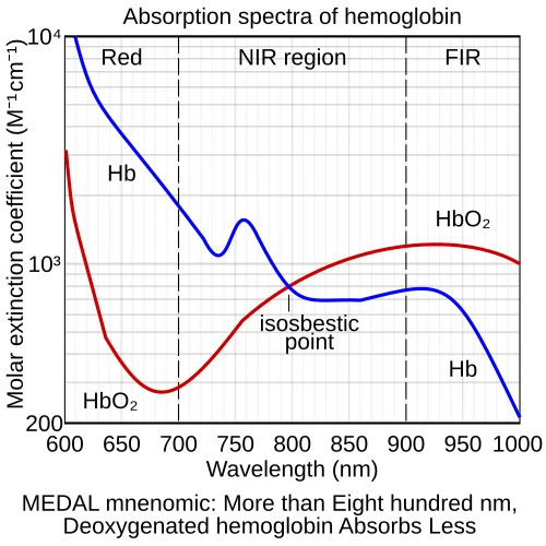
Oxygenated hemoglobin (HbO₂) preferentially absorbs at wavelengths above ~810 nm; deoxygenated hemoglobin (HbR) is the stronger absorber below that crossover point. By illuminating the scalp with light at two wavelengths straddling that boundary and measuring how much comes back, a fNIRS system can estimate the concentration of each species separately using the modified Beer-Lambert law. 2
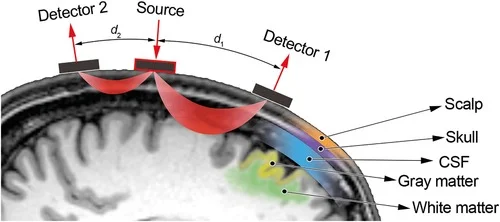
The emitter and detector sit side-by-side on the scalp, separated by 30–35 mm in adult studies (smaller in infants). The light doesn't travel in a straight line — it scatters through tissue in a banana-shaped arc, dipping to a depth roughly half the source-detector separation. Move the optodes further apart and you sample deeper cortex; squeeze them closer and you pick up mostly scalp signal. This geometry determines both what fNIRS can see and its central limitation: subcortical structures, deeper white matter, the hippocampus — all outside reach.
The hemodynamic signal and its relation to fMRI
When a cortical region activates, metabolic demand rises. The brain responds with functional hyperemia: regional blood flow overshoots the oxygen demand, flooding the area with freshly oxygenated blood. HbO₂ rises; HbR falls. This neurovascular coupling — the same mechanism fMRI exploits via the BOLD signal — is what fNIRS measures.
The two signals are closely related but not identical. fNIRS detects both HbO₂ and HbR independently, while BOLD is a composite dominated by HbR changes (deoxygenated hemoglobin is paramagnetic and distorts the local magnetic field). In simultaneous fMRI-fNIRS recordings, the signals correlate well in primary sensory cortex; they diverge more in higher association areas, partly because fNIRS surface sensitivity and fMRI deep sensitivity sample different vascular compartments. 3
What fNIRS uniquely provides is the direction of both signals. A rising HbO₂ alongside a falling HbR is the canonical activation pattern. Seeing HbO₂ rise without a corresponding HbR drop can signal artifactual signal from the scalp (cardiac pulsatility, scalp vasodilation) — a confound that careful paradigm design and short-separation channels can correct for.
What fNIRS opened up that fMRI couldn't
The clearest win has been developmental cognitive neuroscience. Infants cannot hold still in an MRI bore. They can, however, wear a soft optical cap and sit in a caregiver's lap. Within a decade of fNIRS reaching labs, researchers had mapped face-selective responses in the infant occipital cortex, tracked the development of prefrontal working memory representations, and documented how language-selective responses in the perisylvian cortex emerge before infants produce their first words. 1
A second domain is social and naturalistic neuroscience. Most of what we call social cognition — reading a conversation partner's expressions, negotiating, forming impressions — requires interacting with another person in real time. fMRI mostly prohibits this. fNIRS enables it. Hyperscanning studies, where two participants wear fNIRS caps simultaneously, have measured inter-brain synchrony during cooperative tasks, face-to-face communication, and teacher-student instruction — finding that prefrontal coherence between participants predicts behavioral coordination. 4
Third: clinical and applied settings where MRI access is impossible. Neonatal intensive care units use continuous-wave NIRS to monitor cerebral oxygenation in premature infants during surgery. fNIRS studies of schizophrenia have consistently found blunted prefrontal HbO₂ responses during verbal fluency tasks — a pattern now investigated as a potential biomarker. Brain-computer interface work has used fNIRS motor cortex signals to drive assistive devices for people with paralysis.
Advantages and honest limitations
| Dimension | fNIRS | fMRI | EEG |
|---|---|---|---|
| Spatial resolution | ~1–2 cm (cortical surface) | ~2–3 mm (whole brain) | ~cm-scale, source-localization uncertain |
| Temporal resolution | ~10 Hz (hemodynamic lag ~6 s) | ~1 Hz (hemodynamic lag ~6 s) | ms-scale |
| Depth | Cortical surface only | Whole brain | No direct depth |
| Movement tolerance | High | Very low | Moderate |
| Portability | Wearable, wireless options | Fixed, large bore | Portable |
| Cost | Low–moderate | High | Low |
| Simultaneous participants | Yes (hyperscanning) | Impractical | Yes |
The hemodynamic lag is fNIRS's shared weakness with fMRI: both methods track blood flow, which rises and falls over ~6–10 seconds after a neural event. Rapid event-related designs work but sacrifice signal-to-noise. For millisecond-level temporal dynamics — the kind of question best answered by EEG — fNIRS is the wrong tool.
The other major limitation is the shallow depth constraint. This is fundamental, not a hardware problem waiting to be engineered away. No matter how many channels a system has, near-infrared light cannot reach the hippocampus, amygdala, striatum, or thalamus through intact skull in a meaningful way. For research questions requiring those structures, fNIRS is not a substitute for fMRI; it is a complement.
The short-separation channel problem — and its solution
Scalp hemodynamics are real and substantial. When you exercise, blush, or concentrate, blood flow in the scalp changes. For decades this was a significant confound in fNIRS data: you couldn't easily separate a genuine cortical response from a scalp artifact.
The solution was elegant: add a second, short-separation detector about 8 mm from the source. Light at this range barely penetrates the skull — it samples mostly scalp. Subtracting this signal from the long-separation channel removes the scalp component and leaves a purer estimate of cortical hemodynamics. 5 This technical advance, now standard in high-quality fNIRS systems, substantially improved signal specificity and helped resolve earlier replication problems in the literature.
The landmark study: Villringer et al., 1993
The canonical early demonstration of cognitive fNIRS came from Villringer, Planck, Hock, Schleinkofer, and Dirnagl (1993), who used a two-channel near-infrared system to record hemoglobin changes over the motor and sensory cortex during finger tapping in awake adults. 2
The result was clear: contralateral motor cortex showed a rise in HbO₂ and a fall in HbR during movement, peaking around 4–6 seconds after onset and returning to baseline within 10–15 seconds. This matched the BOLD fMRI response profile. It established that the hemodynamic response to cortical activation was optically measurable non-invasively through the intact skull — and that the modified Beer-Lambert law, adapted for tissue scattering by Delpy and colleagues, could extract quantitative concentration changes from the returning light.
That result seems modest now, but it set the template for everything that followed: a functional neuroimaging technique that could be brought outside the scanner room and placed on any head.
Where fNIRS sits in the MIT 9.13 curriculum
MIT 9.13 (The Human Brain, Prof. Nancy Kanwisher) covers fMRI, EEG, TMS, and lesion methods as the canonical toolkit. fNIRS is the newer addition — occupying a position between fMRI (hemodynamic, good spatial resolution, restricted environment) and EEG (electrical, good temporal resolution, high portability). The method is directly relevant to the course's treatment of research methodology and neurovascular coupling, and is increasingly cited in developmental and social cognition literatures that intersect with the face processing, language, and theory of mind modules.
Its rise also tracks the course's broader intellectual concern: what can we actually know about the human brain from the outside? fNIRS is an answer to that question that works in nurseries, in living rooms, and in two-person conversations — contexts where the question of what the human brain does is often most interesting to ask.
Landmark paper: Villringer, A., Planck, J., Hock, C., Schleinkofer, L., & Dirnagl, U. (1993). Near infrared spectroscopy (NIRS): a new tool to study hemodynamic changes during activation of brain function in human adults. Neuroscience Letters, 154(1–2), 101–104. 2
Course connection: MIT 9.13 — Research Methods module; Neurovascular Coupling; Developmental and Social Cognitive Neuroscience.
围绕这条内容继续补充观点或上下文。